Decompression Pars Defect
The Bonati Decompression Pars Defect Procedure relieves pressure on the spinal cord or spinal nerves caused by a fracture of the pars inter-articularis, known as a pars defect.
The spinal canal is formed by a series of bony “arches” that create a tunnel when lined up. The walls of each arch are formed by columns of bones called pedicles. The roof of each arch is formed by a section of bone called the lamina. Each lamina attaches to the one below it through joints on each side called the facet joints. The pars inter-articularis is a bone disconnection between the upper, or superior facet and the lower, or inferior facet through which the nerve passes. This disconnect forms the foramen or opening for the nerve root which exits the spinal canal.

A pars defect, or pars fracture, is a break in the pars inter-articularis, which can lead to a separation of the upper, front portion of the vertebra from the lower, back portion. This may cause compression on nerve roots exiting through the intervertebral foramen, resulting in pain and/or radiculopathy (radiating pain, numbness or weakness in arms, fingers, legs or feet). This defect is commonly associated with spondylolisthesis, which affects the alignment of the vertebrae. For example, if the spondylolisthesis is at L4/L5, the L4 vertebral body will have slipped forward in relation to the L5 vertebral body. If the L4 vertebral body slips backward this is referred to as retrolysthesis which is a less likely alignment resulting from a pars defect. If a vertebrae slips too much, it can press on a nerve and, depending on the level, pain to the lower extremities causing possible muscle weakness. This condition may lead to lower back pain, with radiation into the hips, buttocks or legs.
A decompressive laminectomy is a surgical procedure performed to remove part of the lamina, the vertebral bone that covers the spinal canal. This results in enlargement of the spinal canal and relieves pressure on the spinal nerves or on the spinal cord.
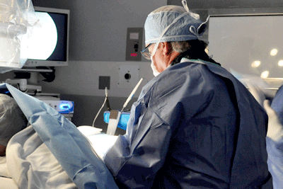
The Bonati Decompression Pars Defect Procedure does not use general anesthesia. Through the use of local anesthesia and conscious IV sedation the patient is comfortable, responsive, and able to provide feedback to the doctors throughout the procedure. This allows our surgeons to target the source of pain with pinpoint accuracy. While in the operating room, the surgical team will confirm the patient is able to complete a series of mobility exercises and verify that the pain has been successfully treated. After the procedure, the patient is transferred to the post-operative care unit for rest and observation, and then a post-operative consultation with the surgeon will help determine if additional procedures included in the surgical plan are necessary. Follow-up surgeries are usually scheduled within a few days of the first surgery, to allow any swelling to subside. During this time, the patient will be given a regimen of walking therapy.
The Bonati Spine Institute offers a comprehensive review of MRI studies or MRI reports to prospective patients at no obligation to the patient. One of our surgeons will review your MRI scan and note each condition, its cause and severity. Then, we will discuss the findings with you to see if you are a candidate for the Patented Bonati Spine Procedures.
Contact The Bonati Spine Institute today and speak with a Patient Advocate to answer your questions.
The Bonati Spine Institute offers MRI or CT Scan reviews for potential patients suffering from chronic pain which can be caused by a variety of spine problems. A surgeon will provide feedback and possible treatment options, for suitable Bonati Spine Procedures candidates. Request your MRI Review now.
